
Goldstein, Bob

December 14, 2023

We are interested in understanding how cells develop into organisms. We love the nematode C. elegans, because it allows us to readily combine a great number of useful techniques, including techniques of cell biology, direct manipulation of cells, forward and reverse genetics, biochemistry, molecular biology, biophysics, and live imaging of cells and their dynamic, cytoskeletal components. Current work in the lab addresses several fundamental questions in cell and developmental biology — how cells move to specific positions during development, how cells change shape, how developmental patterning mechanisms tell cell biological mechanisms what to do where and when, how intercellular signals act to polarize cells, and how the mitotic spindle is positioned in cells.

We have also been developing a relative of C. elegans and Drosophila, a water bear (tardigrade), to study how developmental mechanisms can evolve to produce organisms with different forms and how biological materials can survive unusual extremes.

C. elegans and tardigrades

Dowen, Rob

March 28, 2019

Research Synopsis:

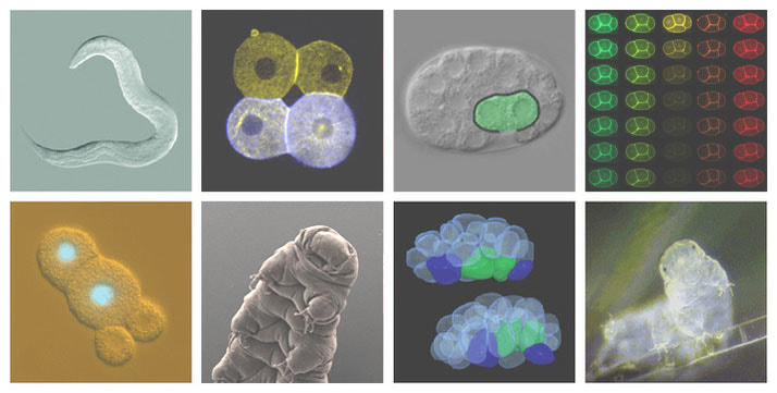
Appropriate allocation of cellular lipid stores is paramount to maintaining organismal energy homeostasis and is coordinated by a network of multi-tissue endocrine signals. Dysregulation of these pathways can manifest in human metabolic syndromes, including cardiovascular disease, obesity, diabetes, and cancer. The goal of my lab is to elucidate the molecular mechanisms that govern the storage, metabolism, and intercellular transport of lipids; as well as understand how these circuits interface with other cellular homeostatic pathways (e.g., growth and aging). We utilize C. elegans as a model system to interrogate these evolutionarily conserved pathways, combining genetic approaches (forward and reverse genetic screens, CRISPR) with genomic methodologies (ChIP-Seq, mRNA-Seq, DNA-Seq) to identify new components and mechanisms of metabolic regulation.

Recent Publications:

Dowen, RH. CEH-60/PBX and UNC-62/MEIS coordinate a metabolic switch that supports reproduction in C. elegansDevelopmental Cell 2019 Apr 22;49(2):235-50. PMID: 30956009

Dowen, RH, Breen, PC, Tullius, T, Conery, AL, Ruvkun, G. A microRNA program in the C. elegans hypodermis couples to intestinal mTORC2/PQM-1 signaling to modulate fat transport. Genes & Development 2016 Jul 1;30(13):1515-28. PMID: 27401555

Riedel, CG, Dowen, RH, Lourenco, GF, Kirienko, NV, Heimbucher, T, West, JA, Bowman, SK, Kingston, RE, Dillin, A, Asara, JM, and Ruvkun, G. DAF-16 employs the chromatin remodeller SWI/SNF to promote stress resistance and longevity. Nature Cell Biology 2013 May;15(5):491-501. PMID: 23604319

Dowen, RH, Pelizzola, M, Schmitz, RJ, Lister, R, Dowen, JM, Nery, JR, Dixon, JE, and Ecker, JR. Widespread dynamic DNA methylation in response to biotic stress. Proc Natl Acad Sci USA 2012 Aug 7;109(32):E2183-91. PMID: 22733782

Lister, R*, Pelizzola, M*, Dowen, RH, Hawkins, RD, Hon, G, Tonti-Filippini, J, Nery, JR, Lee, L, Ye, Z, Ngo, Q, Edsall, L, Antosiewicz-Bourget, J, Stewart, R, Ruotti, V, Millar, AH, Thomson, JA, Ren, B, and Ecker, JR. Human DNA methylomes at base resolution show widespread epigenomic differences. Nature 2009 Nov 19;462(7271):315-322. *Equal contribution. PMID: 19829295

Shiau, Celia

June 27, 2016

Macrophages are highly dynamic and widespread blood cells that play many important functions in vertebrates. They are the main phagocytes throughout the body, responsible for clearing away dying cells, damaged tissue, and pathogens, to maintain tissue integrity. Macrophages circulate in the bloodstream as monocytes or are stationed in strategic locations of the body as tissue macrophages where their phagocytic roles are critical, such as microglia in the brain, Kupffer cells in the liver, Langerhans cells in the skin, and osteoclasts in the bone.

Of particular interest are the normal roles and mechanisms of macrophages and microglia in the development and maintenance of the nervous system that remain far less understood than their functions in disease and injury. In the healthy brain, microglia have been implicated in shaping brain circuitry and neuronal development as well as in possibly affecting behavioral outcomes. They have unique embryonic origins from primitive macrophages that migrate into the brain and remain thereafter through life. These versatile glial cells provide the first line of defense and respond to a wide variety of environmental factors, such as protein aggregates, apoptotic cells, injured tissue, and pathogens, as well as to intracellular dysfunctions. Overall, the developmental process by which macrophages take residence and differentiate into tissue macrophages, and the contribution of macrophages to normal animal development remain not well understood.  We are addressing these two fundamental areas of macrophage biology in the context of how macrophages participate in the nervous system.


Nimchuk, Zachary

July 24, 2015

Plants contain discrete populations of self-renewing stem cells that give rise to the diverse differentiated cell types found throughout the plant. Stem cell function is therefore ultimately responsible for the aesthetic and economic benefits plants provide us. My lab uses multiple approaches to dissect plant stem cell signaling networks including genetics, genomics, live tissue imaging, and cell biological and biochemical methods.  

McKay, Daniel J.

July 1, 2014

Research in the lab focuses on how a single genome gives rise to a variety of cell types and body parts during development. We use Drosophila as a model organism to investigate (1) how transcription factors access DNA to regulate complex patterns of gene expression, and (2) how post-translational modification of histones contributes to maintenance of gene expression programs over time. We combine genomic approaches (e.g. chromatin immunoprecipitation followed by high-throughput sequencing) with Drosophila genetics and transgenesis to address both of these questions. Defects in cell fate specification and maintenance of cell identity often occur in human diseases, including cancer. (website)

McKay_research_image

Rogers, Steve

June 24, 2011

The research in our lab is centered on understanding the mechanisms and principles of cellular movement. Cytoskeletal filaments – composed of actin and microtubules – serve as a structural scaffolding that defines the architecture of the cytoplasm and gives cells the ability to divide, crawl, and change their shapes. We are interested in understanding how cells regulate cytoskeletal dynamics to produce motility. Our primary model system is the fruit fly, Drosophila melanogaster as it allows us to use functional genomic tools and classical genetic techniques to study gene function at the level of individual cells and during development. Current projects in the lab address mechanisms of microtubule dynamic instability, crosstalk between the actin and microtubule cytoskeletal networks, and the regulation of cellular contractility during Drosophila gastrulation.

Kieber, Joseph

June 20, 2011

Hormones influence virtually every aspect of plant growth and development. The elucidation of the molecular mechanisms controlling the biosynthesis and perception of these hormonal signals, and how these signals are integrated with each other and with other developmental and environmental signals remain fundamental questions in plant biology. There are three main areas of focus to the research in my laboratory: cytokinin signaling, the regulation of ethylene biosynthesis, and the regulation of cell elongation. Using a combination of genetic, molecular and biochemical approaches we are attempting to elucidate elements involved in these processes and ultimately to understand how these elements contribute to control plant growth and development.

Cytokinin Signal Transduction

Cytokinins were discovered by their property of promoting cell division. These N6-substituted adenine-based molecules have been associated with various developmental roles including germination, shoot and root development and leaf senescence. A model for cytokinin signal transduction has emerged that is similar to bacterial two-component systems (Fig. 1). Two-component elements in Arabidopsis are encoded by multi-gene families and similar families have been identified in the monocots maize and rice.

Figure 1: General model of cytokinin signaling. See: To and Kieber (2008), Trends in Plant Science, for details.

We seek to understand how these signaling elements interact with each other, how this pathway outputs to regulate the many processes that cytokinin regulates and to use mutants in various signaling components to further define the role of cytokinin in plant growth and development.

Regulation of Ethylene Biosynthesis

The simple gas ethylene has been recognized as a plant hormone since the turn of the century. It has been shown to influence a diverse array of plant processes, such as leaf and flower senescence and abscission, fruit ripening and the response to a wide variety of stresses. In order to understand how ethylene or any signaling molecule affects development, one must consider how its biosynthesis is controlled and the molecular mechanisms underlying its perception. Almost all plant tissues have the capacity to make ethylene, although in most cases the amount of ethylene produced is very low. There is a diverse group of factors that increase the level of ethylene biosynthesis, including other hormones (auxin, cytokinin, ethylene), numerous stresses, as well as various developmental events. We have taken a genetic approach to understanding how ethylene biosynthesis is regulated. Two classes of mutants have been isolated: those that overproduce ethylene (Eto mutants) and mutants that fail to increase ethylene biosynthesis in response to a specific inducer, cytokinin (Cin mutants). Together, these mutants identify elements involved in regulating ethylene biosynthesis. Studies from our lab have provided compelling evidence that ACS protein stability is regulated and have converged on a common mechanism. Research in our lab focuses on unraveling this mechanism regulating ACS protein stability. We are exploring the role of protein phosphorylation in this process. We are examining if the stability of ACS protein increases during developmental events that are associated with a rise in ethylene biosynthesis or in response to exogenous cues such as light. We are using various genetic screens to identify novel elements involved in controlling ACS protein stability. These studies will shed light on the mechanism regulating the regulation of the stability of ACC synthase proteins and how this contributes to the control of the biosynthesis of ethylene.

Regulation of Cell Expansion

The regulation of cell expansion is a primary determinant in the size and shape of plant organs. Understanding how cells regulate this process is crucial in understanding the development of plant form. The Arabidopsis root is an excellent model system to dissect this process as it has a relatively simple, well defined architecture and mutants that alter cell elongation can easily be identified by their effect on root length. In the root, two distinct regions of cell expansion can be distinguished. Both longitudinal and radial cell expansion occurs in the root apical meristem, defining the root diameter and moving cells into the elongation zone of the root. In the elongation zone, just above the meristem, elongation rates are also uniform, but are much higher and occur almost exclusively in the longitudinal direction. This polar cell expansion is known as anisotropy. Both the extent and orientation of cell expansion is regulated in plants. Cell expansion can be a dynamic process, and the orientation of expansion can change in response to various stimuli, such as wounding and hormonal treatments. Expansion of cells is driven by turgor pressure and the re

lative alignment and composition of cell wall material, which determines both the extent and orientation of elongation.

Figure 2: Phenotype of WT and AIK mutant seedlings. Note that the aik mutant root swells as a result of isotropic expansion of the root cells.

The plant cell wall is comprised of cellulose microfibrils that are crosslinked with glycans and are associated with a pectin matrix and various extracellular proteins. The primary load bearing elements of the cell wall are the cellulose microfibrils, and thus their orientation and crosslinking are key factors in both the direction and extent of cell expansion.In anisotropically elongating cells, the cellulose microfibrils are wound in a helical spiral transversely around the cell, in a manner that has been likened to hoops around a barrel. This arrangement allows expansion of the cell specifically in the longitudinal direction by stretching of this cellulose “spring”, but restricts expansion in the transverse or radial direction. Cellulose microfibrils are synthesized at the plasma membrane by a hexameric protein complex called the terminal complex or rosette, and the polysaccharides made by this complex are extruded into the extracellular space through some type of a pore in the plasma membrane, where they then associate with other cellulose chains to form microfibrils.

A major question regarding cell expansion is how is the deposition of the cellulose microfibrils is regulated to give, for example, primarily transverse microfibrils in root cells. Early studies revealed that the cytoplasmic microtubules were aligned transversely in some plant cells, correlated to the orientation of the cellulose microfibrils in those cells, and this and other data led to the idea that the microtubules act as a template for the synthesis of the cellulose microfibrils.

Our lab seeks to explore the links between ethylene, cell expansion and a pair of receptor-like kinase gene that we have evidence may link these processes. We identified a number a member of the leucine-rich receptor like Ser/Thr protein kinase (LRR-RLK) family in Arabidopsis (AIK1) that interacts with ACC synthase. Disruption of AIK results in Arabidopsis results in a plant in which the root cells lose their ability to elongation anisotropically. Further analysis suggests that ACC may act as a novel signal in this process. Molecular and genetic analysis has identified additional components in what now defines a novel regulatory circuit that controls the extend and orientation of cell expansion. These studies will shed light on how plants regulate both the extent and orientation of cell expansion and will help understand how the plant cell wall is constructed.

Peifer, Mark

June 14, 2011

Peifer Lab Website

Follow our lab on Facebook

Teaching   |   Undergraduate Research

Developmental Biology at UNC  |  Cytoskeletal research at UNC

Cell adhesion, cytoskeletal regulation and Wnt signaling

in development and cancer


Biomedical science has twin goals; to explain the many amazing properties of our own bodies and those of other animals, and to use this information to reveal the causes of disease and to suggest possible treatments. We work at the interface between cell and developmental biology, focusing on the epithelial tissues that form the basic architectural unit of our bodies and of those of other animals.  We explore how the machinery mediating cell adhesion, cytoskeletal regulation and Wnt signaling regulates cell fate and tissue architecture in development and disease.  Epithelial tissues like skin, lung, colon, and breast are affected in many cancers. Cancer results from alterations in normal cell behaviors. To explore underlying causes of epithelial tumors, we need to understand the basic cellular machinery that links cell adhesion, signal transduction and cytoskeletal regulation during normal development.

We focus on the machinery that modulates cell-cell adhesion and connects cell junctions to the actin cytoskeleton, thus shaping the architecture of epithelial tissues.  We also explore the machinery that transduces and regulates Wnt signaling, which helps determine cell fates.  Wnt signaling is inappropriately activated in colon and other cancers, while the cell adhesion machinery is inactivated in most metastatic tumors.  We study these processes in the fruit fly Drosophila, combining classical and modern genetic tools with state-of-the-art cell biology, microscopy, and biochemistry,  thus capitalizing on the speed of this model system and its synergy with vertebrate cell biology, and supplement this with work on cultured Drosophila cells and mammalian cells.  Like all good science, our work sometimes leads us in unexpected directions–our recent wor on the roles of centrosomes in genome stability is an example.

Wnt signals are one of the five signal transduction pathways that shape virtually all cell fates and which are inappropriately activated in most solid tumors.  The key regulated effector of Wnt signaling is the protein ß-catenin.  Wnt signaling acts by regulating its stability.  In the absence of Wnt signaling, ß-catenin is targeted for proteasomal destruction by a multi-protein complex called the destruction complex.  In the presence of Wnt signals, the destruction complex is inactivated, and ß-catenin levels rise, allowing it to enter the nucleus and work with TCF proteins to regulate Wnt target genes.  In our lab, we seek to determine how the tumor suppressor APC, a key component of the destruction complex, regulates both Wnt signaling and the cytoskeleton.  We use both the fruit fly Drosophila and cultured human colon culture cells to unravel the mechanisms by which APC works.  We combine powerful genetic tools and state of the art microscopy.  We are currently exploring how APC regulates assembly and disassembly of the destruction complex as part of a catalytic cycle.  We are also exploring separate roles APC plays in regulating the cytoskeleton and thus ensures high fidelity chromosome segregation, and branching off from this the interplay between mitotic fidelity regulators, checkpoints and apoptosis which maintains genome stability. Finally, we explore novel biological roles for Wnt signaling during development.


Figure 3. Armadillo-GFP localization to cell-cell junctions during dorsal closure in a living embryo.
In studying adhesion, our challenge is to alter our static model of adhesion to explain the remarkable cellular events of morphogenesis that shape the embryonic body plan and build tissues and organs. To do so, we must understand the dynamic regulation of cell adhesion and how it is coordinated with the cytoskeleton. We visualize these processes via state-of-the art confocal microscopy and live-imaging, using fluorescently-tagged versions of adhesion and cytoskeletal regulatory proteins, as well as probes that allow us to visualize the actin and microtubule cytoskeletons. This allows us to examine cell behavior and the cell biological events underlying it during dynamic events of morphogenesis, such as dorsal closure.  In searching for regulators of adhesion and the cytoskeleton, we have focused on the non-receptor tyrosine kinase Abelson (Abl). Mutations in Abl cause two forms of human leukemia. We found that Abl coordinately regulates adhesion and the dynamics of the actin cytoskeleton. We are currently exploring the mechanisms by which Abl regulates complex events of morphogenesis.  We also are exploring the functions of proteins that directly regulate actin dynamics, including Diaphanous-class formins and Enabled/VASP proteins, some of which are targets of Abl.  In parallel, we are examining proteins that help form the dynamic links between the cadherin-catenin complex and the actomyosin cytoskeleton.   We focus on the small GTPases Rap1 and the junctional protein Canoe/Afadin.  In addition to their role in adhesion, these proteins also regulate cell polarity, and we are actively pursuing their roles in both apical-basal and planar polarity.


To learn more about our work, visit our wpcf-lab-website via the link above, where you can meet the people in the lab and learn more about their work. It’s an exciting time to be working at the interface between cell and developmental biology, and we are always looking for talented and enthusiastic graduate students and postdocs to add to our group.  You can also follow us on Facebook or check out our videos on Vimeo.

Obtaining Armadillo Antibody

The anti-Armadillo antibody is now available from the Developmental Studies Hybridoma Bank, an NIH funded facility that produces antibodies for the research community at cost. They will sell you anti-Armadillo 7A1 mouse monoclonal antibody at $10/ml. You can reach them by phone at 319-335-3826 or by wpcf-emailfaculty at dshb@uiowa.edu. Perhaps the easiest way to reach them is at their home page at http://dshb.biology.uiowa.edu/.

If you need more information about the use of the antibody, feel free to contact us. We use it at 1:40 in situ on embryos, 1:20 for immunoprecipitations, and at 1:400 on Westerns.

Good luck with your experiments.


Figure 4. Armadillo protein is normally found in the adherens junctions surrounding each cell. However, in cells which have received Wingless signal, Armadillo protein also accumulates in the cytoplasm and the nucleus, where we suspect it may be involved in activating transcription of target genes. Panel A shows an embryo double labeled with anti-Armadillo antibody (red) and anti-Engrailed antibody (green). Engrailed is a transcription factor and marks the nucleus. Some nuclei are yellow, showing co-localization of Armadillo and Engrailed in the nuclei of cells receiving Wingless signal. Panels B and C are the single labeled images.