The research in our lab is centered on understanding the mechanisms and principles of cellular movement. Cytoskeletal filaments – composed of actin and microtubules – serve as a structural scaffolding that defines the architecture of the cytoplasm and gives cells the ability to divide, crawl, and change their shapes. We are interested in understanding how cells regulate cytoskeletal dynamics to produce motility. Our primary model system is the fruit fly, Drosophila melanogaster as it allows us to use functional genomic tools and classical genetic techniques to study gene function at the level of individual cells and during development. Current projects in the lab address mechanisms of microtubule dynamic instability, crosstalk between the actin and microtubule cytoskeletal networks, and the regulation of cellular contractility during Drosophila gastrulation.
Combining genetics with real-time analysis of living cells by video or digital enhanced microscopy to examine the role of dynein and other microtubule motor proteins in spindle and nuclear dynamics.
Using specific chromosomal or cytoskeletal perturbations to understand cell cycle control of late mitotic events.
Synopsis
Figure 1. Spindles are longer in histone depleted cells (kinetochores in green, SPBs in red).
Accurate chromosome segregation at each cell division is one of the most fundamental processes in a living organism. This complex process requires the coordination of cytoskeletal components such as microtubules and microtubule motor proteins that form the mitotic spindle with the replication and attachment of chromosomes to the segregation apparatus. Both the high fidelity of chromosome segregation and genome integrity are essential for normal development and cellular propagation. The ease of genetic and molecular manipulations to address the mechanism of chromosome segregation n the context of a live cell makes the budding yeast an excellent model system to address the mechanism of spindle assembly and the maintenance of genome integrity in mitosis. Our lab takes a broad approach to understanding mitosis in yeast. Recent projects in the lab have addressed the role of plus end microtubule binding proteins such as Bim1 and microtubule motor proteins on spindle integrity and the contribution of pericentric chromatin to regulating spindle length.
The site of microtubule attachment to the chromosome is the kinetochore, a complex of over 60 proteins assembled at a specific site on the chromosome, the centromere. Almost every kinetochore protein identified in yeast is conserved throughout phylogeny and the organization of the kinetochore in yeast may serve as the fundamental unit of attachment for mammalian cells. More recently we have become interested in the role of two different classes of ATP binding proteins, cohesions (Smc3, Scc1) and chromatin remodeling factors (Cac1, Hir1, Rdh54), in the structural organization of the kinetochore and how these classes of proteins contribute to the fidelity of chromosome segregation.
Chromosomes are compacted into increasingly complex chromatin structures within eukaryotic nuclei. High-throughput sequence-based assays have been developed to identify regions of nucleosome-depleted open chromatin that mark all types of regulatory elements genome-wide in tissues and cell-types. The computational integration of these data with related gene expression, transcription factor binding, and epigenetic data provide a more complete picture of the complex process of gene transcription and regulation. With these data, we are also investigating the effects of genetic variation on regulation, as can been seen through allelic imbalance in signal from chromatin and transcription factor data, as well as in quantitative trait loci (QTL)-based analyses of these data across individuals. Our computational biology lab both applies established analysis tools to data generated in collaboration with other labs as well as develops new analytical techniques to make novel associations.
Inflammatory Bowel Disease
Inflammatory bowel disease (IBD), primarily consisting of Crohn’s disease and ulcerative colitis, is the result of an inappropriate immune response to the intestinal microbiota in a genetically susceptible individual. We have partnered with Dr. Shehzad Sheikh, MD, PhD (Dept of Medicine, CGIBD) to uncover molecular and microbial characteristics of IBD disease phenotypes. We hypothesize that changes in the chromatin landscape in key intestinal cell types such as macrophages, are influenced by the host genetic background and significantly contribute to aberrant intestinal inflammation. Using both human tissue and mouse models, we seek to identify where chromatin is altered, the impacts on gene expression, the contributions of genetic variation, and the relationship to the microbial community in tissues and cells from affected individuals.
Environmental Toxicogenomics
Exposure to naturally occurring toxicants or by-products of manufacturing process can result in serious health challenges. We hypothesize that toxicant exposure can alter normal cellular function through changes in chromatin architecture and transcriptional profiles in tissues or cells contributing to the onset of medical complications. In a collaboration with Dr. Samir Kelada, PhD (Dept of Genetics), we are investigating the effects of exposure to ozone on the lung inflammatory response in the genetically diverse Collaborative Cross mouse model resource. In particular, we are assaying how ozone-induced chromatin structure and gene expression changes in alveolar macrophages, along with genetic variation, explain variability in ozone response.
Hormones influence virtually every aspect of plant growth and development. The elucidation of the molecular mechanisms controlling the biosynthesis and perception of these hormonal signals, and how these signals are integrated with each other and with other developmental and environmental signals remain fundamental questions in plant biology. There are three main areas of focus to the research in my laboratory: cytokinin signaling, the regulation of ethylene biosynthesis, and the regulation of cell elongation. Using a combination of genetic, molecular and biochemical approaches we are attempting to elucidate elements involved in these processes and ultimately to understand how these elements contribute to control plant growth and development.
Cytokinin Signal Transduction
Cytokinins were discovered by their property of promoting cell division. These N6-substituted adenine-based molecules have been associated with various developmental roles including germination, shoot and root development and leaf senescence. A model for cytokinin signal transduction has emerged that is similar to bacterial two-component systems (Fig. 1). Two-component elements in Arabidopsis are encoded by multi-gene families and similar families have been identified in the monocots maize and rice.
Figure 1: General model of cytokinin signaling. See: To and Kieber (2008), Trends in Plant Science, for details.
We seek to understand how these signaling elements interact with each other, how this pathway outputs to regulate the many processes that cytokinin regulates and to use mutants in various signaling components to further define the role of cytokinin in plant growth and development.
Regulation of Ethylene Biosynthesis
The simple gas ethylene has been recognized as a plant hormone since the turn of the century. It has been shown to influence a diverse array of plant processes, such as leaf and flower senescence and abscission, fruit ripening and the response to a wide variety of stresses. In order to understand how ethylene or any signaling molecule affects development, one must consider how its biosynthesis is controlled and the molecular mechanisms underlying its perception. Almost all plant tissues have the capacity to make ethylene, although in most cases the amount of ethylene produced is very low. There is a diverse group of factors that increase the level of ethylene biosynthesis, including other hormones (auxin, cytokinin, ethylene), numerous stresses, as well as various developmental events. We have taken a genetic approach to understanding how ethylene biosynthesis is regulated. Two classes of mutants have been isolated: those that overproduce ethylene (Eto mutants) and mutants that fail to increase ethylene biosynthesis in response to a specific inducer, cytokinin (Cin mutants). Together, these mutants identify elements involved in regulating ethylene biosynthesis. Studies from our lab have provided compelling evidence that ACS protein stability is regulated and have converged on a common mechanism. Research in our lab focuses on unraveling this mechanism regulating ACS protein stability. We are exploring the role of protein phosphorylation in this process. We are examining if the stability of ACS protein increases during developmental events that are associated with a rise in ethylene biosynthesis or in response to exogenous cues such as light. We are using various genetic screens to identify novel elements involved in controlling ACS protein stability. These studies will shed light on the mechanism regulating the regulation of the stability of ACC synthase proteins and how this contributes to the control of the biosynthesis of ethylene.
Regulation of Cell Expansion
The regulation of cell expansion is a primary determinant in the size and shape of plant organs. Understanding how cells regulate this process is crucial in understanding the development of plant form. The Arabidopsis root is an excellent model system to dissect this process as it has a relatively simple, well defined architecture and mutants that alter cell elongation can easily be identified by their effect on root length. In the root, two distinct regions of cell expansion can be distinguished. Both longitudinal and radial cell expansion occurs in the root apical meristem, defining the root diameter and moving cells into the elongation zone of the root. In the elongation zone, just above the meristem, elongation rates are also uniform, but are much higher and occur almost exclusively in the longitudinal direction. This polar cell expansion is known as anisotropy. Both the extent and orientation of cell expansion is regulated in plants. Cell expansion can be a dynamic process, and the orientation of expansion can change in response to various stimuli, such as wounding and hormonal treatments. Expansion of cells is driven by turgor pressure and the re
lative alignment and composition of cell wall material, which determines both the extent and orientation of elongation.
Figure 2: Phenotype of WT and AIK mutant seedlings. Note that the aik mutant root swells as a result of isotropic expansion of the root cells.
The plant cell wall is comprised of cellulose microfibrils that are crosslinked with glycans and are associated with a pectin matrix and various extracellular proteins. The primary load bearing elements of the cell wall are the cellulose microfibrils, and thus their orientation and crosslinking are key factors in both the direction and extent of cell expansion.In anisotropically elongating cells, the cellulose microfibrils are wound in a helical spiral transversely around the cell, in a manner that has been likened to hoops around a barrel. This arrangement allows expansion of the cell specifically in the longitudinal direction by stretching of this cellulose “spring”, but restricts expansion in the transverse or radial direction. Cellulose microfibrils are synthesized at the plasma membrane by a hexameric protein complex called the terminal complex or rosette, and the polysaccharides made by this complex are extruded into the extracellular space through some type of a pore in the plasma membrane, where they then associate with other cellulose chains to form microfibrils.
A major question regarding cell expansion is how is the deposition of the cellulose microfibrils is regulated to give, for example, primarily transverse microfibrils in root cells. Early studies revealed that the cytoplasmic microtubules were aligned transversely in some plant cells, correlated to the orientation of the cellulose microfibrils in those cells, and this and other data led to the idea that the microtubules act as a template for the synthesis of the cellulose microfibrils.
Our lab seeks to explore the links between ethylene, cell expansion and a pair of receptor-like kinase gene that we have evidence may link these processes. We identified a number a member of the leucine-rich receptor like Ser/Thr protein kinase (LRR-RLK) family in Arabidopsis (AIK1) that interacts with ACC synthase. Disruption of AIK results in Arabidopsis results in a plant in which the root cells lose their ability to elongation anisotropically. Further analysis suggests that ACC may act as a novel signal in this process. Molecular and genetic analysis has identified additional components in what now defines a novel regulatory circuit that controls the extend and orientation of cell expansion. These studies will shed light on how plants regulate both the extent and orientation of cell expansion and will help understand how the plant cell wall is constructed.
DNA damage is a constant challenge to cells and a primary event leading to cancer. Double-strand breaks – when both strands of the double helix are severed – is among the most lethal forms of damage; it is essential that such breaks be repaired efficiently and with high fidelity. We seek to understand the molecular mechanisms through which this is achieved. We use primarily genetic approaches (classical and molecular, forward and reverse), using the fruit fly Drosophila melanogaster as a model organism.
Double-strand breaks occur spontaneously due to problems during DNA replication, or they can be induced by exposure to ionizing radiation or clastogenic chemicals. Meiotic cells actually induce programmed double-strand breaks. The goal of meiosis is to generate haploid gametes from a diploid cell. To do this, homologous chromosomes must pair and stick together until anaphase of meiosis I. How do they stick together? By making crossovers. The pathway for generating crossovers begins with the formation of double-strand breaks. These breaks are repaired in a highly regulated way to ensure that crossovers are generated in the right places, but not in the wrong places. We are interested in the molecular mechanisms of this specialized repair process and how it differs from double-strand break repair in non-meiotic cells.
These projects are discussed in more detail on our lab web site.
Cell adhesion, cytoskeletal regulation and Wnt signaling
in development and cancer
Biomedical science has twin goals; to explain the many amazing properties of our own bodies and those of other animals, and to use this information to reveal the causes of disease and to suggest possible treatments. We work at the interface between cell and developmental biology, focusing on the epithelial tissues that form the basic architectural unit of our bodies and of those of other animals. We explore how the machinery mediating cell adhesion, cytoskeletal regulation and Wnt signaling regulates cell fate and tissue architecture in development and disease. Epithelial tissues like skin, lung, colon, and breast are affected in many cancers. Cancer results from alterations in normal cell behaviors. To explore underlying causes of epithelial tumors, we need to understand the basic cellular machinery that links cell adhesion, signal transduction and cytoskeletal regulation during normal development.
We focus on the machinery that modulates cell-cell adhesion and connects cell junctions to the actin cytoskeleton, thus shaping the architecture of epithelial tissues. We also explore the machinery that transduces and regulates Wnt signaling, which helps determine cell fates. Wnt signaling is inappropriately activated in colon and other cancers, while the cell adhesion machinery is inactivated in most metastatic tumors. We study these processes in the fruit fly Drosophila, combining classical and modern genetic tools with state-of-the-art cell biology, microscopy, and biochemistry, thus capitalizing on the speed of this model system and its synergy with vertebrate cell biology, and supplement this with work on cultured Drosophila cells and mammalian cells. Like all good science, our work sometimes leads us in unexpected directions–our recent wor on the roles of centrosomes in genome stability is an example.
Wnt signals are one of the five signal transduction pathways that shape virtually all cell fates and which are inappropriately activated in most solid tumors. The key regulated effector of Wnt signaling is the protein ß-catenin. Wnt signaling acts by regulating its stability. In the absence of Wnt signaling, ß-catenin is targeted for proteasomal destruction by a multi-protein complex called the destruction complex. In the presence of Wnt signals, the destruction complex is inactivated, and ß-catenin levels rise, allowing it to enter the nucleus and work with TCF proteins to regulate Wnt target genes. In our lab, we seek to determine how the tumor suppressor APC, a key component of the destruction complex, regulates both Wnt signaling and the cytoskeleton. We use both the fruit fly Drosophila and cultured human colon culture cells to unravel the mechanisms by which APC works. We combine powerful genetic tools and state of the art microscopy. We are currently exploring how APC regulates assembly and disassembly of the destruction complex as part of a catalytic cycle. We are also exploring separate roles APC plays in regulating the cytoskeleton and thus ensures high fidelity chromosome segregation, and branching off from this the interplay between mitotic fidelity regulators, checkpoints and apoptosis which maintains genome stability. Finally, we explore novel biological roles for Wnt signaling during development.
Figure 3. Armadillo-GFP localization to cell-cell junctions during dorsal closure in a living embryo.
In studying adhesion, our challenge is to alter our static model of adhesion to explain the remarkable cellular events of morphogenesis that shape the embryonic body plan and build tissues and organs. To do so, we must understand the dynamic regulation of cell adhesion and how it is coordinated with the cytoskeleton. We visualize these processes via state-of-the art confocal microscopy and live-imaging, using fluorescently-tagged versions of adhesion and cytoskeletal regulatory proteins, as well as probes that allow us to visualize the actin and microtubule cytoskeletons. This allows us to examine cell behavior and the cell biological events underlying it during dynamic events of morphogenesis, such as dorsal closure. In searching for regulators of adhesion and the cytoskeleton, we have focused on the non-receptor tyrosine kinase Abelson (Abl). Mutations in Abl cause two forms of human leukemia. We found that Abl coordinately regulates adhesion and the dynamics of the actin cytoskeleton. We are currently exploring the mechanisms by which Abl regulates complex events of morphogenesis. We also are exploring the functions of proteins that directly regulate actin dynamics, including Diaphanous-class formins and Enabled/VASP proteins, some of which are targets of Abl. In parallel, we are examining proteins that help form the dynamic links between the cadherin-catenin complex and the actomyosin cytoskeleton. We focus on the small GTPases Rap1 and the junctional protein Canoe/Afadin. In addition to their role in adhesion, these proteins also regulate cell polarity, and we are actively pursuing their roles in both apical-basal and planar polarity.
To learn more about our work, visit our wpcf-lab-website via the link above, where you can meet the people in the lab and learn more about their work. It’s an exciting time to be working at the interface between cell and developmental biology, and we are always looking for talented and enthusiastic graduate students and postdocs to add to our group. You can also follow us on Facebook or check out our videos on Vimeo.
Obtaining Armadillo Antibody
The anti-Armadillo antibody is now available from the Developmental Studies Hybridoma Bank, an NIH funded facility that produces antibodies for the research community at cost. They will sell you anti-Armadillo 7A1 mouse monoclonal antibody at $10/ml. You can reach them by phone at 319-335-3826 or by wpcf-emailfaculty at dshb@uiowa.edu. Perhaps the easiest way to reach them is at their home page at http://dshb.biology.uiowa.edu/.
If you need more information about the use of the antibody, feel free to contact us. We use it at 1:40 in situ on embryos, 1:20 for immunoprecipitations, and at 1:400 on Westerns.
Good luck with your experiments.
Figure 4. Armadillo protein is normally found in the adherens junctions surrounding each cell. However, in cells which have received Wingless signal, Armadillo protein also accumulates in the cytoplasm and the nucleus, where we suspect it may be involved in activating transcription of target genes. Panel A shows an embryo double labeled with anti-Armadillo antibody (red) and anti-Engrailed antibody (green). Engrailed is a transcription factor and marks the nucleus. Some nuclei are yellow, showing co-localization of Armadillo and Engrailed in the nuclei of cells receiving Wingless signal. Panels B and C are the single labeled images.
My research interests are focused on the regulation of gene activity in animal cells, in particular regulation of gene expression during the cell cycle by postranscriptional mechanisms. One system we study is the regulation of histone mRNA, both during the mammalian cell cycle and during early development in frogs and sea urchins. Histone mRNAs are the only mRNAs that do not have polyA tails, ending instead in a conserved stem-loop structure. Histone mRNAs are present only in S-phase cells and most of the regulation is mediated by the 3′ end of histone mRNA. Histone genes lack introns, and histone mRNA is formed by a single processing reaction, cleavage to form the 3′ end of the histone mRNA. The mRNA is then immediately transported to the cytoplasm. Both the cleavage reaction to form histone mRNA and the half-life of the histone mRNA are regulated during the cell cycle. We have cloned the cDNA for the stem-loop binding protein (SLBP) that binds the 3′ end of histone mRNA and participates in all aspects of histone mRNA metabolism. SLBP is a critical factor involved in regulating histone mRNA levels. Our current interests are in understanding how SLBP carries out its multiple functions; RNA binding, 3′ processing, transport, stimulator of translation and regulating histone mRNA half-life. In addition, we are studying how SLBP itself is regulated and how this regulation connects the other cell cycle regulators with the regulation of histone mRNA.
Sea Urchin
In embryos which undergo a very rapid series of cell divisions after fertilization, there is an exponentially increasing demand for histones to assemble the newly replicated DNA into chromatin. During this time the histone mRNAs are not cell-cycle regulated but are stable for multiple cell cycles. We have cloned embryo specific SLBPs from these stages and are determining how they function to regulate histone mRNA metabolism in frog and sea urchin embryos. We are also studying the role of the G1 cyclins, cyclin D and cyclin E in the regulation of these early cell cycles that lack gap phases.
The research in our laboratory interrogates fundamental molecular, cellular and developmental biological processes. In particular, we are interested in the roles played by small ribonucleoproteins (RNPs) in a genetic disease, called Spinal Muscular Atrophy. We are also focused on the functions of histone post-translational modifications (PTMs) in the regulation of eukaryotic gene expression, important for understanding disease mechanisms in many different types of cancer.
How do intracellular innate immune receptors of the NLR family function?
How is the plant associated microbiome condition plant growth and environmental response?
And how do these commensal microbes navigate or evade the plant immune system?
Synopsis
Many interactions between plants and microbes begin with specific recognition. The nature of this recognition, and the interpretation of subsequent signal transduction by both plant and microbe have profound impact on the outcome of the interaction. Plants have evolved effective mechanisms to recognize pathogenic microbes and halt their biotrophic or necrotrophic growth in the plant. Active plant defense mechanisms obviously force the selection of microbe variants which can evade the plant’s recognition capabilities. This evolutionary tug of war has led to a complex set of both plant and microbe genes, whose interactions lead to a successful plant resistance reaction. As well as a potentially large array of cognitive gene functions, a number of subsequent signal transduction steps must be necessary to generate a completely effective resistant phenotype. Plant-microbe interactions can also benefit the plant and plant’s select a small and taxonomically constrained set of microbes from the very rich microbiome of the soil. These commensals help the plant access minerals and can protect against pathogens. We study both the molecular mechanisms of the plant immune system and the intricacies of how that immune system sculpts the well organized and functional root microbiome. Our ultimate aim is to use knowledge, genetics and microbes from nature to enhance plant performance and soil sustainability across the globe by defining fundamental rules of microbiome assembly and function.
My lab has studied the genetics of plant-pathogen interactions since 1989. Our two main interests are the control of pathogen recognition by plants via their two tiered immune system consisting of extracellular pattern recognition receptors and intracellular nucleotide-binding, leucine-rich repeat (NLR) receptors, and the formation and function of the root microbiome. In the immune system, we study NLR activation and its outcomes, activation of transcriptional re-programming to result in pathogen growth restriction and, ultimately, hypersensitive cell death. We also study the structure, function and evolutionary genomics of bacterial pathogen type III effectors and oomycete effectors. These pathogen proteins suppress host defense by manipulating host protein machinery. We are dedicated to mapping that set of host machines using the evolved tools of the pathogen to identify them. We study these interactions at the molecular and structural level. In the root microbiome, we are defining the community structure of plant rhizoplane and endophytic microbiomes and trying to define design rules for small bacterial consortia that will enhance plant health and productivity. This project relies on genomics, ecological modeling, metabolic modeling and both forward and reverse genetics. We have expertise and momentum in these research arenas and we collaborate widely to extend our technical and intellectual reach. My lab is a diverse set of individuals from several countries who each bring different expertise to their projects, and to the other projects in the group. We stress small team approaches to the problems introduced above. My students and post-docs thus have access to a variety of inputs about their work. We work on biological scales from nanometers to small mesocosms. Our lab alumnae have been successful in job placement in a variety of careers.
Arabidopsis thaliana
We use the reference model plant species, Arabidopsis thaliana, in our research.
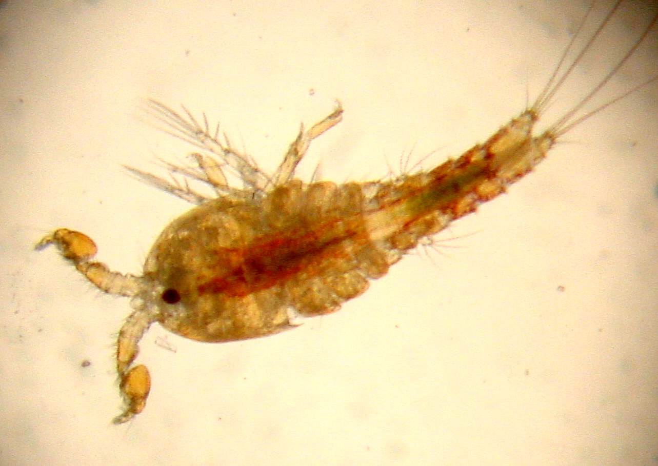
My research focuses on the evolutionary genetics and genomics of aspects of speciation and adaptation. The basic concepts I study are how one species can split into two over time and what factors influence how organisms adapt to their environment. Current work centers on exploring questions in these areas using copepods and moths as model systems. Here are some interests:
Speciation Genetics: Genetic basis of hybrid breakdown in copepods and the appearance of reproductive isolation
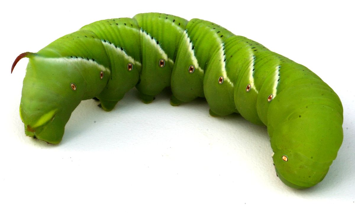
Genetic basis of physiological adaptation: Temperature and salinity tolerance in copepods and the moth Manduca sexta
Comparative genomics and using next generation sequencing to study the genetic basis of speciation and adaptation
Population genetics and molecular sequence evolution
Conservation genetics: Outbreeding depression, population genetics of threatened species
Synopsis
My research addresses the nature of genetic variation that underlies speciation and adaptation. Specifically, I attempt to unravel how genetic changes at the molecular level can lead to phenotypic changes of evolutionary significance. A major thrust of my research program has been to understand how genetic variation within populations translates into variation between populations and species, and to determine the impact of natural selection on this process. In my current work I am targeting specific genetic systems and using genome-wide approaches to determine the regions of the genome that could be involved in generating reproductive isolation that occurs after mating (postzygotic reproductive isolation). I am also examining the physiological and fitness consequences of genetic variation using both targeted gene and genome-wide approaches. Specifically we are looking at the evolution of temperature and salinity adaptation across populations. My work to date has largely focused on two primary systems systems-copepods and moths.
Copepods:
The harpacticoid copepod Tigriopus californicus inhabits rocky, intertidal splash pools in a patchy distribution along the west coast of North America. Populations of this species display dramatic genetic differentiation even between relatively proximate localities. Crosses between these populations typically show hybrid breakdown (decreases in fitness of F2 individuals). Understanding the genetic basis of this hybrid breakdown could yield insights into the early stages of the process of speciation (link). Recent work in the lab has also focused on the nature of adaptation in thermal tolerance among populations of this species and its implications for reproductive isolation in this system. This work could help reveal how species will be able to handle future changes in temperature environments (link).
Moths:
My lab is now working in collaboration with Joel Kingsolver’s lab in a project studying thermal biology and genetics in caterpillars of Manduca sexta, the tobacco hornworm. This project focuses on how organisms respond to variation in temperature over time (i.e. warmer during the day and cooler at night) and will help us understand better how to predict how organisms will be able to handle a changing climate. We will be focusing in my lab in how the caterpillars are responding on the genetic and protein levels and how this can help us understand what is happening at the level of the whole organism. Another interesting part of the work is comparing field and lab-adapted animals to which can illuminate how thermal tolerance can change over relatively short periods of time (link).
I have also worked in the past on the evolution of pheromone communication systems. This work largely focused on the evolution of one protein, the pheromone-binding protein, and how it contributes to discrimination by male moths and differentiation among species (link).
EDUCATION
B.S. in Zoology, Michigan State University, 1993
Ph.D. in Genetics and Development, Cornell University, 1999